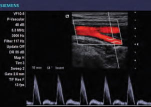
Ultrasounds are a safe imaging technique used to view the movement and structure of internal organs and/or blood vessels in real time. Also called sonograms, ultrasound images are captured using high-frequency sound waves, not radiation. The sound waves’ echoes are recorded and displayed in real-time, visual images.
Ultrasound technicians use a hand-held device called a transducer to send sound waves through the body. The ultrasound travels through fluids and soft tissue, but if it hits denser material, it bounces back or echoes. It’s these echoes that create the images. Dense objects cause more ultrasound to bounce back and appear darker in the image produced. Various shades of gray in the images represent different densities. An ultrasound examination is fast, with very little discomfort. No radiation or X-rays are necessary. Ultrasound procedures are relatively quick from start to finish.