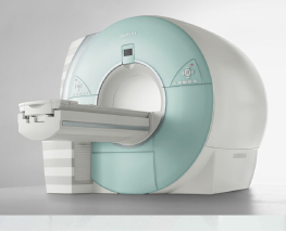
University Center Imaging is equipped with the Siemens MAGNETOM High-Field 1.5T. We offer several different types of MRI procedures including:
MRI Scans – High-Field 1.5T Short Bore, MRI Scans: The “bore” refers to the opening (tube) of the MRI scanner. A wide bore MRI is shorter and wider, that combines expansive patient comfort with no compromise to image quality or scan power. This scanner is the industry standard and allows Radiologists to obtain detailed images of the body’s internal anatomy without exposing you to radiation. Exams range from 15 to 60 minutes depending on the type of exam.
MRCP Scans: Magnetic resonance cholangiopancreatography is a specialized MRI focused on obtaining images of the liver, bile ducts, gallbladder, pancreas and pancreatic duct. It’s generally used to diagnose abdominal pain. The entire exam takes about 45 minutes from start to finish.
MRI Breast: Using the same state-of-the-art equipment as our other services, this MRI looks specifically at the breast and offers valuable information about many breast conditions by compiling hundreds of images, all cross-sectional in all three directions that cannot be obtained through mammography or ultrasound. The MRI produces no radiation and the procedure has no known hazards to your health.
MRI Angiography: The display of blood vessels known as MR angiography is an accurate, non-invasive means of obtaining information about arteries of the head, neck and body. MRI is a painless, non-invasive procedure, uses no radiation and has no known side effects.
Your physician may recommend an MRI scan for a variety of reasons. This non-invasive tool gives them a highly detailed look inside the human body for examining the following:


MRI examinations take about 15 to 60 minutes from start to finish. Your doctor may order your MRI to include contrast, a fluid injected into the vein to make certain details appear clearer in the images produced.
When you enter the MRI suite, you will be asked to lay on the examination table on your back, either headfirst or feet first depending on the type of scan performed. Our goal is to ease any anxiety and help you feel relaxed during the procedure. You can listen to music or your favorite CD during the exam. If you need to, you can even have a family member in the room. The table slides into the MRI machine’s opening, and when the scan commences, you will hear the hum of the device. This is all normal. A microphone allows you to speak to your technologist throughout the procedure and the technologist will speak to you. However, during the scan, you must remain as still as possible.

Save time! To expedite your process, complete your Registration online through our Patient Portal.

Once your scan is complete, a Radiologist will examine the images. Your physician will receive the images and reports via PACS and fax. Contact your physician directly to discuss your results. Results are also available through the Patient Portal. Allow 3 to 5 business days.